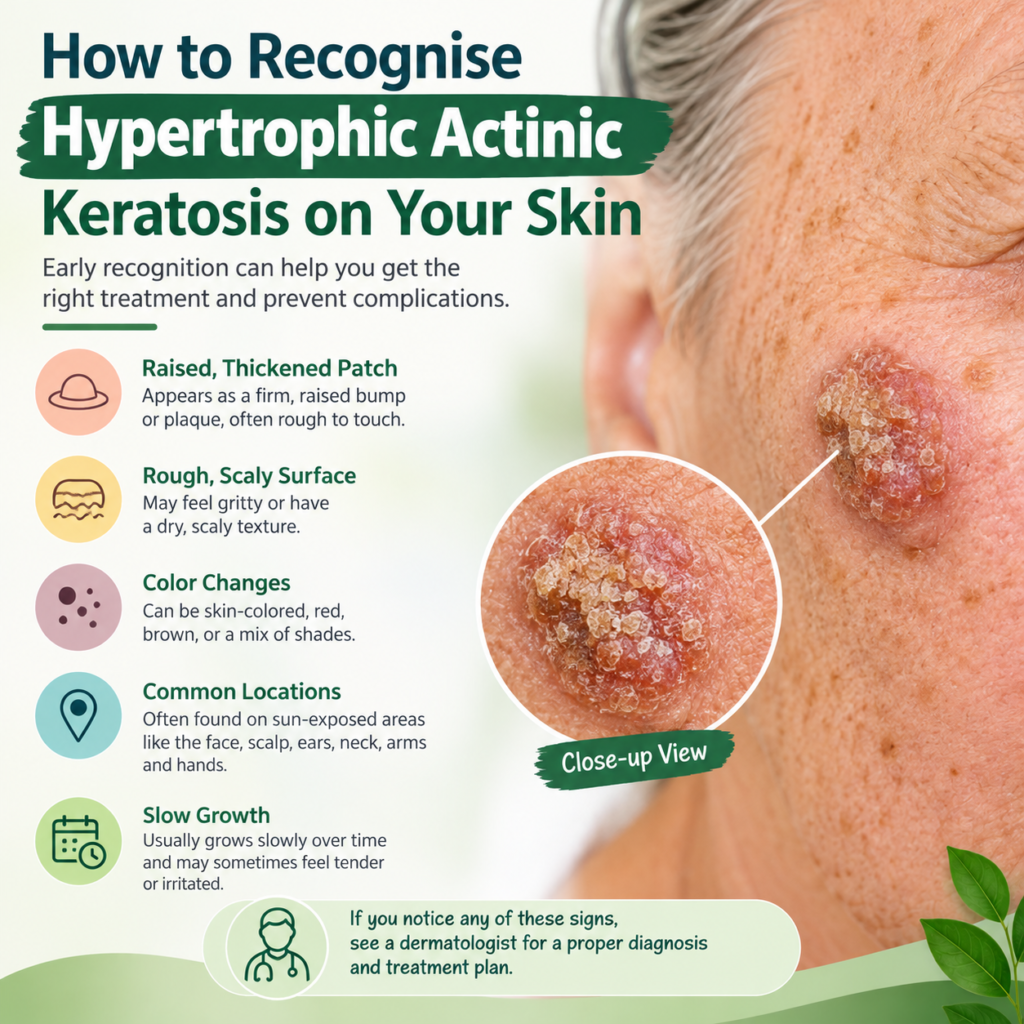
Hypertrophic Actinic Keratosis can appear as a thick, rough, scaly growth that requires professional assessment.
Hypertrophic Actinic Keratosis is a thickened, rough skin lesion caused by long-term ultraviolet (UV) exposure. It often develops on sun-exposed areas, including the face, scalp, ears, hands, and forearms. Recognising its appearance early matters because some lesions can resemble other skin conditions, including skin cancers.
Although these growths are not always cancerous, they should never be ignored. Learning about their texture, colour, location, and changes over time can help you recognise warning signs and seek appropriate medical advice. This guide explains what to look for, which symptoms deserve attention, how dermatologists diagnose these lesions, and what treatment options may help protect your skin.
What Is Hypertrophic Actinic Keratosis?
Hypertrophic Actinic Keratosis is a thicker, often more prominent form of actinic keratosis. Actinic keratoses develop when repeated UV exposure damages the DNA of skin cells, particularly keratinocytes, which form much of the skin’s outer layer.
The hypertrophic form produces excess keratin, a protein that helps protect the skin. As keratin accumulates, the affected area can become raised, hardened, scaly, or crusted. Some lesions feel like sandpaper, while others develop a horn-like projection.
These growths are commonly found in people with a history of frequent sun exposure, especially those with fair skin or extensive sun damage. However, people of other skin tones can also develop actinic keratoses.
Actinic keratosis is considered a precancerous skin condition because some lesions may progress to squamous cell carcinoma, a type of skin cancer. A thick or horn-like appearance does not prove that cancer is present, but it makes professional assessment particularly important.
How to Identify the Main Skin Changes
The appearance of a lesion can vary depending on its thickness, colour, and location. Checking your skin in good lighting may help you notice changes that deserve medical attention.
1. Thick, raised growths
One of the most recognisable features is a raised patch with a hard, uneven surface. The lesion may look like a small wart or a compact mound of dead skin. Unlike a flat sunspot, it often has noticeable thickness.
A thick lesion may catch on clothing, feel uncomfortable during washing, or become irritated when rubbed. However, these features alone cannot confirm the diagnosis.
2. Rough, scaly texture
The surface often feels dry, gritty, or sandpaper-like. You might notice white, yellowish, or brown scales that cling firmly to the affected skin.
Some growths develop a crusted surface that returns after the crust falls away. Repeated crusting, particularly when accompanied by bleeding or tenderness, should prompt a medical examination.
3. Changes in colour
The affected area may appear pink, red, brown, grey, yellowish, or similar to your natural skin tone. Colour alone is not a reliable way to distinguish a harmless growth from a precancerous lesion.
In some cases, surrounding skin also shows signs of sun damage, including uneven pigmentation, fine wrinkles, and several rough patches.
4. A horn-like projection
A particularly thick lesion may develop a pointed, keratin-filled projection known as a cutaneous horn. This resembles a tiny animal horn growing from the skin.
A horn is a physical description rather than a diagnosis. Its base may contain an actinic keratosis, a benign growth, or a skin cancer. A dermatologist should assess it, and a biopsy may be necessary.
Where Do These Lesions Usually Appear?
9
Sun exposure largely determines where these lesions develop. Areas that receive repeated sunlight are especially vulnerable.
Common locations
- Face: The forehead, temples, nose, and cheeks may develop rough or raised patches.
- Scalp: People with thinning hair or bald scalps may notice thick, crusted spots.
- Ears: The rims and outer surfaces are frequently exposed to sunlight.
- Hands: The backs of the hands and fingers can develop rough growths.
- Forearms: These areas receive considerable UV exposure during outdoor activities.
- Lips: Persistent roughness or scaling on the lower lip may indicate actinic cheilitis, a related sun-damage condition.
The location can provide an important clue, but it does not establish the diagnosis. A new, thickened growth in any location should be examined if it persists or changes.
What Causes These Skin Growths?
The main cause is cumulative exposure to ultraviolet radiation from sunlight or artificial tanning devices. UV rays damage the genetic material of skin cells, and repeated exposure can lead to abnormal cell growth.
Several factors can increase the likelihood of developing these lesions.
Long-term sun exposure
Spending years outdoors without adequate protection allows UV damage to accumulate. Outdoor workers, gardeners, farmers, and people who regularly participate in outdoor sports may face greater exposure.
Sunburns can contribute to skin damage, but repeated exposure can also be harmful without obvious burning.
Age and skin characteristics
The risk generally increases with age because damage builds up over time. People with fair skin, freckles, light-coloured eyes, or a history of sunburn may be more susceptible.
Nevertheless, darker skin tones do not eliminate the possibility of actinic keratosis. Everyone can benefit from sun protection and regular skin checks.
Weakened immune function
People taking immunosuppressive medicines or living with certain conditions that weaken immune function may develop more lesions and face a higher risk of skin cancer.
Previous skin cancer and extensive sun damage also warrant closer monitoring. If you have these risk factors, ask your clinician how often you should receive a skin examination.
How Is It Different from Other Skin Conditions?
Several skin conditions can resemble a thick precancerous lesion. Trying to identify the exact cause by appearance alone may delay appropriate treatment.
| Skin condition | Typical appearance | Important distinction |
| Common actinic keratosis | Rough, scaly, often relatively thin patch | Usually has less pronounced thickening |
| Seborrhoeic keratosis | Waxy, brown or tan growth that appears stuck on | Usually benign, although it can resemble other lesions |
| Common wart | Firm, rough bump, sometimes with tiny dark dots | Often has a characteristic wart-like surface |
| Squamous cell carcinoma | Persistent, firm, scaly or crusted growth | May enlarge, ulcerate, become painful or bleed |
| Basal cell carcinoma | Pearly bump or persistent sore | May bleed easily or fail to heal |
These descriptions are general clues, not a substitute for examination. Some lesions overlap in appearance, and a thick keratotic growth may be difficult to distinguish from early squamous cell carcinoma.
Warning Signs You Should Never Ignore
A longstanding rough spot deserves attention if its behaviour changes. Although irritation does not automatically mean cancer, certain developments require prompt assessment.
Contact a dermatologist if you notice:
- Rapid enlargement or a sudden increase in thickness.
- Persistent pain, tenderness, or sensitivity.
- Repeated bleeding without a clear injury.
- An open sore that does not heal.
- Ulceration, persistent crusting, or discharge.
- A hard base or a growth that feels fixed to the underlying skin.
- A lesion that changes significantly in shape, colour, or texture.
These symptoms can occur with different skin conditions, including squamous cell carcinoma. Arrange an urgent medical assessment if a lesion grows quickly, bleeds repeatedly, or becomes painful. Do not wait for every warning sign to appear before seeking advice.
How Do Dermatologists Diagnose It?
A dermatologist will examine the lesion and ask when it first appeared, whether it has changed, and whether you have a history of sun damage or skin cancer.
Skin examination
The clinician assesses the lesion’s thickness, surface, borders, colour, and location. They may examine the surrounding skin for additional precancerous spots or signs of other skin cancers.
Dermoscopy
A handheld dermatoscope magnifies the skin and reveals structures that are difficult to see with the naked eye. This can help the clinician assess suspicious features, although it cannot always provide a definitive answer.
Skin biopsy
A biopsy may be recommended when the lesion is unusually thick, painful, ulcerated, bleeding, or suspicious for invasive cancer. It is particularly important when a thick keratotic growth cannot be confidently distinguished from squamous cell carcinoma.
During this procedure, the clinician removes a sample or the entire lesion, depending on the circumstances. A laboratory examines the tissue under a microscope to establish the diagnosis.
If you suspect Hypertrophic Actinic Keratosis, avoid scraping the surface or attempting to remove the growth yourself. A professional assessment helps determine whether further investigation is needed.
Medical Treatments That May Be Recommended
Treatment depends on the lesion’s thickness, location, number, and diagnostic certainty. The dermatologist will also consider your medical history and whether cancer needs to be excluded.
Cryotherapy
Cryotherapy uses liquid nitrogen to freeze and destroy the abnormal skin cells. It is commonly used for individual actinic keratoses.
The treated area may blister, crust, or become temporarily red before healing. Very thick lesions may require a different approach or additional treatment.
Curettage
Curettage involves scraping away the abnormal tissue with a specialised instrument. It may be suitable for particularly thick lesions.
The removed tissue can be sent for laboratory examination when clinically indicated. If cancer is suspected, the clinician will select an appropriate diagnostic and treatment procedure.
Prescription creams
For suitable cases, prescription medicines such as fluorouracil, imiquimod, diclofenac, or tirbanibulin may be recommended. These treatments have different indications, schedules, and side effects.
They are often used for multiple lesions or broader areas of sun-damaged skin. They are not interchangeable, and the appropriate choice depends on the clinical situation.
Photodynamic therapy
This treatment uses a light-sensitive medication followed by a specific light source to destroy abnormal cells. It may be useful when multiple lesions affect a particular area.
Treatment can cause redness, discomfort, and temporary sensitivity to light. Your clinician will explain the preparation and aftercare required.
When Hypertrophic Actinic Keratosis is exceptionally thick or suspicious, the priority is to establish the diagnosis and exclude invasive cancer rather than simply applying a cream.
Can Natural Approaches Help Protect Your Skin?
People often seek a Natural Treatment for Actinic Keratosis because they prefer gentle skincare or want to reduce further sun damage. However, no home remedy has been established as a reliable way to remove a thick precancerous lesion or rule out cancer.
Supportive skincare can complement medical care, but it should not replace examination or recommended treatment.
Everyday skin protection
Consider these practical habits:
- Apply broad-spectrum sunscreen with SPF 30 or higher to exposed skin.
- Reapply sunscreen approximately every two hours outdoors and after swimming or heavy sweating.
- Wear a wide-brimmed hat, long sleeves, and protective clothing.
- Seek shade during periods of strong sunlight.
- Avoid tanning beds and deliberate tanning.
- Use a gentle, fragrance-free moisturiser if your skin feels dry.
These measures help reduce further UV damage and the development of additional lesions. They cannot reverse every existing abnormality, so continue attending recommended dermatology appointments.
What About Herbal Skincare and Supplements?
Interest in Natural Remedies for Actinic Keratosis often centres on plant extracts, antioxidant-rich foods, moisturisers, and supplements. Although some ingredients may support general skin health, evidence that they can eliminate an established precancerous lesion is insufficient.
Avoid applying concentrated essential oils, acidic household ingredients, abrasive scrubs, or unverified herbal pastes to a suspicious growth. These products may cause irritation, burns, or delayed diagnosis.
Choosing Products with Professional Guidance
If you use Herbal Care Products or other botanical supplements, review the ingredients with your healthcare professional, particularly if you take medicines or have ongoing health conditions. Supplements should not be presented as a substitute for proven dermatological treatment.
A balanced diet containing vegetables, fruit, whole grains, and adequate protein supports general wellbeing. However, no particular food or supplement can guarantee protection from skin cancer.
The safest approach combines evidence-based treatment, sensible skincare, and regular monitoring. Discuss complementary products with your dermatologist before using them on a lesion that has not been diagnosed.
How to Monitor Your Skin at Home
Regular skin checks can help you identify new growths and changes in existing lesions. Choose a well-lit room and use a mirror to inspect areas that are difficult to see.
Check your face, scalp, ears, neck, hands, forearms, and other sun-exposed areas. Ask someone you trust to help examine your scalp or back if necessary.
When you find a suspicious spot, record its location and the date you first noticed it. A photograph may help you track visible changes, but it cannot establish whether the growth is cancerous.
Pay particular attention to lesions that become thicker, develop a horn-like projection, or repeatedly form a crust. Do not pick at them to see what lies underneath.
If you have previously been diagnosed with actinic keratoses, follow your dermatologist’s recommended schedule for follow-up examinations. New lesions may develop even after successful treatment of existing ones.
Frequently Asked Questions
Is a thick actinic keratosis always cancerous?
No. It is a precancerous lesion, not automatically an invasive cancer. However, thick growths can resemble squamous cell carcinoma, so a dermatologist may recommend a biopsy to clarify the diagnosis.
Can a lesion disappear without treatment?
Some actinic keratoses may regress temporarily or disappear, but others persist or return. Spontaneous improvement does not guarantee that the underlying risk has disappeared. Seek medical advice about any persistent or suspicious growth.
Does it hurt?
Many lesions cause no symptoms. Others itch, sting, feel tender, or become irritated by clothing. Persistent pain, rapid growth, or repeated bleeding deserves prompt assessment.
How quickly should I seek medical advice?
Arrange an appointment when you discover a persistent, thick, rough lesion that could be actinic keratosis. Seek more urgent assessment if it grows rapidly, ulcerates, becomes painful, or bleeds repeatedly.
Can sunscreen treat an existing lesion?
Sunscreen does not reliably remove an established lesion. Its main role is to limit further UV damage and reduce the risk of additional actinic keratoses. Medical treatment may still be necessary.
Protect Your Skin Through Early Action
Recognising suspicious changes is an important step towards protecting your long-term skin health. A thick, rough, scaly, or horn-like growth on a sun-exposed area deserves attention, especially if it changes, hurts, or bleeds.
Although Hypertrophic Actinic Keratosis is not synonymous with skin cancer, its appearance can overlap with more serious conditions. A dermatologist can assess the lesion, arrange a biopsy when needed, and recommend appropriate treatment.
Protect your skin with regular sunscreen use, protective clothing, and routine examinations. Most importantly, do not rely on appearance or home remedies to decide whether a suspicious growth is harmless. Early professional assessment offers the clearest path to an accurate diagnosis and appropriate care.